Conserved allomorphs of MR1 drive the specificity of MR1-restricted TCRs
Overview
This study investigates the specificity of the MR1-restricted TCR from clone MC.7.G5. Results show recognition is restricted to the MR1*04 allomorph and is not cancer-specific, reacting to healthy cells expressing MR1*04. G-Rex® systems were used to seed and expand CD3+ T cells during the transduction process.
What this paper found
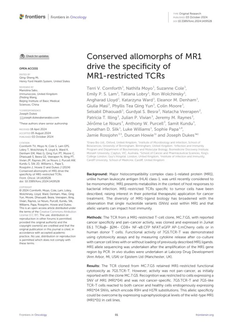
A TCR cloned from the MR1-restricted clone MC.7.G5 and expressed in human T cells as 7G5.TCR-T did not show pan-cancer activity; it recognized cells carrying the rare MR1*04 allele, including healthy cells. Only nine of 133 cancer cell lines (7%) gave IFN-γ above 50 pg/ml, and responses to MR1*04-bearing targets were two- to 110-fold greater than to other lines. Monocytes and B cells from MR1*01/*04 donors, but not MR1*01 homozygous donors, triggered IFN-γ and granzyme B release. Overexpressing MR1*01 at supraphysiological levels also drove reactivity, while the bacterial ligand Ac-6-FP abrogated responses to MR1*04 lines. Two 7G5-like TCRs, A4 and C1, behaved similarly, producing IFN-γ and granzyme B against MR1*04 heterozygous B cells and monocytes.
Note: The work combines in vitro assays with mouse xenograft models; the authors note that the ligand(s) bound to MR1 required for 7G5.TCR-T activity remain unidentified.
To our knowledge, this is the first demonstration of an MR1restricted TCR that has MR1 allomorph specificity. — Terri V. Cornforth, Nathifa Moyo, Suzanne Cole, Emily P. S. Lam, Tatiana Lobry, Ron Wolchinsky, Angharad Lloyd, Katarzyna Ward, Eleanor M. Denham, Giulia Masi, Phyllis Tea Qing Yun, Colin Moore, Selsabil Dhaouadi, Gurdyal S. Besra, Natacha Veerapen, Patricia T. Illing, Julian P. Vivian, Jeremy M. Raynes, Jérôme Le Nours, Anthony W. Purcell, Samit Kundu, Jonathan D. Silk, Luke Williams, Sophie Papa, Jamie Rossjohn, Duncan Howie, Joseph Dukes, Frontiers in Oncology
Study details
- Authors
- Terri V. Cornforth, Nathifa Moyo, Suzanne Cole, Emily P. S. Lam, Tatiana Lobry, Ron Wolchinsky, Angharad Lloyd, Katarzyna Ward, Eleanor M. Denham, Giulia Masi, Phyllis Tea Qing Yun, Colin Moore, Selsabil Dhaouadi, Gurdyal S. Besra, Natacha Veerapen, Patricia T. Illing, Julian P. Vivian, Jeremy M. Raynes, Jérôme Le Nours, Anthony W. Purcell, Samit Kundu, Jonathan D. Silk, Luke Williams, Sophie Papa, Jamie Rossjohn, Duncan Howie, Joseph Dukes
- Institution
- Enara Bio Ltd., Oxford, United Kingdom · Institute of Microbiology and Infection, School of Biosciences, University of Birmingham, Birmingham, United Kingdom · Infection and Immunity Program and Department of Biochemistry and Molecular Biology, Biomedicine Discovery Institute, Monash University, Clayton, VIC, Australia · School of Cancer and Pharmaceutical Sciences, King's College London, Guy's Hospital, London, United Kingdom · Institute of Infection and Immunity, Cardiff University, School of Medicine, Cardiff, United Kingdom
- Journal
- Frontiers in Oncology
- Year
- 2024
- G-Rex® model(s)
- G-Rex®10M
- Cell type(s)
- TCR T Cells
- Application area
- Hematologic malignancy · Solid tumor
- Study phase
- Preclinical
How G-Rex® was used
Positively selected CD3+ T cells (15 × 10^6) were seeded with CD3/CD28 Dynabeads in 10M G-Rex® systems in TexMACS with 5% human AB serum and 20 ng/ml IL-2 and incubated overnight, then transduced with lentivirus to express a TCR. After a further 24 h, fresh medium with IL-2 was added to the G-Rex® systems, where cells for some in vivo experiments were treated with CRISPR Cas9 puromycin. CD3+ cells were harvested, beads removed and cells frozen 10 days after transduction and expansion.
Explore related

Full citation
Cornforth TV, Moyo N, Cole S, et al. Conserved allomorphs of MR1 drive the specificity of MR1-restricted TCRs. Front Oncol. 2024;14:1419528.
Building a similar workflow?
Talk to a ScaleReady specialist about how the G-Rex® platform can support your program — from process development to clinical-scale manufacturing.