In vitro efficacy of cell therapy products determined using a G-Rex® co-culture assay
Overview
This note from the G-Rex® manufacturer sets out a killing assay in which effector and tumor cells share a G-Rex® 6-well plate for weeks, with bioluminescence tracking growth and no media change. Anti-CD19 CAR T cells held lymphoma in check until a CD19-negative population emerged after three weeks.
What this paper found
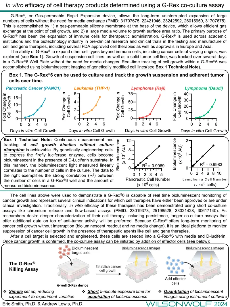
A long-term co-culture killing assay in the G-Rex® 6 Well Plate lets researchers follow tumor cell growth and the effect of cell therapies without media changes. Target cells engineered to express firefly luciferase are seeded with D-luciferin, and bioluminescence, which tracks cell number linearly, is imaged repeatedly. Suspension lines (THP-1, Raji, Daudi) and a solid tumor line (PANC1) grew over several days in this format. In a demonstration, anti-CD19 CAR T cells controlled lymphoma growth, but a CD19-negative population emerged after three weeks of undisturbed culture. The suggested set-up uses 100 μM D-luciferin and 35 mL of medium per well, and the assay was also applied to non-engineered tumor antigen-specific T cells.
Note: The cell lines shown illustrate real-time bioluminescent monitoring of cancer growth; the document presents a method with example data rather than a full efficacy study.
As researchers desire deeper characterization of their cell therapy, including persistence, longer co-culture assays that offer additional data on top of anti-tumor activity will be preferred. — Eric Smith, Andrew Lewis
Study details
- Authors
- Eric Smith, Andrew Lewis
- Year
- 2024
- G-Rex® model(s)
- G-Rex®6 Well Plate
- Cell type(s)
- CAR T Cells
- Application area
- Hematologic malignancy · Solid tumor
- Study phase
- Preclinical
How G-Rex® was used
Firefly luciferase-engineered target cells are seeded into a G-Rex® 6 Well Plate with medium and D-luciferin, and once cancer growth is confirmed, effector cells are added to start the co-culture. Each well is given the recommended 35 mL of medium with 100 μM D-luciferin, and growth is tracked without media changes by repeatedly imaging bioluminescence in a ChemiDoc instrument. Suspension lines (THP-1, Raji, Daudi) and a solid tumor line (PANC1) were followed over several days this way. In the CD19 CAR T example, lymphoma growth was controlled until a CD19-negative population emerged after three weeks of undisturbed culture.
Explore related

Building a similar workflow?
Talk to a ScaleReady specialist about how the G-Rex® platform can support your program — from process development to clinical-scale manufacturing.